Gum Biopsy in Memphis TN
OR CALL (901) 300-4162
In 2021, over 50,000 people in the United States were diagnosed with oral cancer. If not caught early, oral cancer can spread quickly and cause bleeding, swelling, jaw pain, and even fatality. Fortunately, most cases of oral cancer can be resolved if caught soon. One of the most common methods used to diagnose oral cancer is through a gum biopsy.
What is a gum Biopsy?
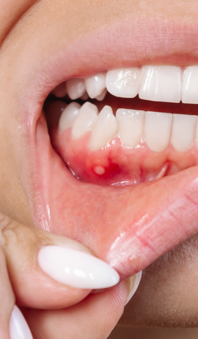
The purpose of a gum biopsy is to identify the source of abnormal gum tissue. If it’s a cancerous growth, it is essential to receive treatment as soon as possible. You should get a gum biopsy if you experience any of the following:
- A sore or lesion that doesn’t go away after two weeks
- A white or red patch or ulcer
- Lumps along the side of the mouth
- Loose teeth or receding gums
- Long-lasting swelling
- Difficulty chewing or swelling
Along with a gum biopsy, a patient may have to receive an x-ray, MRI, or CT scan.
What is the procedue like?
A gum biopsy is a relatively quick procedure that is typically performed by a dentist, periodontist, or oral surgeon. You may be asked to refrain from eating or to reveal any medications you are on prior to the biopsy.
Before the procedure, Dr. Godat or Dr. King will numb your gums by applying a local anesthetic. In more invasive cases, you may be given a general anesthetic that will keep you asleep. Depending on your needs and the condition of your gums, you will receive one of three types of gum biopsies, each of which has its own unique features. Here’s what you can expect from each one.
INCISIONAL BIOPSY
Most patients receive an incisional biopsy, during which a small portion of the abnormal tissue is extracted and then examined. During the incision, you may experience some minor discomfort or bleeding. At the end of the procedure, the incision may be stitched up or sealed using a laser. Typically, stitches are dissoluble and will simply dissolve in about a week. However, in some cases, the stitches will need to be removed by a professional. In this case, you will be asked to return to the periodontist after a week or so.
EXCISIONAL BIOPSY
When a patient has a small, accessible legion, they usually receive an excisional biopsy. In this procedure, that entire legion or growth is removed for examination. The actual procedure is similar to that of an incisional biopsy–after the growth is taken out, the area may be stitched to prevent bleeding.
PERCUTANEOUS BIOPSY
A percutaneous biopsy involves injecting a needle into the skin for testing. There are two kinds of this biopsy:
- Fine needle (used for growths that are highly visible and accessible)
- Core needle (used when a high amount of tissue is needed)
In a fine needle biopsy, the needle is inserted straight into the lesion and used to remove cells. The injection may be repeated several times across the same area.
During a core needle biopsy, a small circular blade is pressed against the abnormal area to form a round mold. A needle is then used to remove a circular portion of the gum, which brings a group of cells that then undergo testing. One of the advantages of core needle biopsies is that they rarely require stitches to heal.
Gum Biopsy Results
Once the tissue sample has been removed, it will be sent to a pathologist who will examine it and provide a prognosis. Potential diagnoses include:
- Oral or mouth cancer
- Benign growth
- Blood disorders
- Systemic disorders
Gum biopsies are useful for more than identifying cancer–they can also be utilized to catch dangerous conditions that affect your bloodstream or bodily systems. One of the most common conditions found through a gum biopsy is systemic amyloidosis, during which abnormal proteins are formed in the organs and distributed throughout the body. Another condition that affects the gums is thrombotic thrombocytopenic purpura, a clotting disorder that prevents blood from reaching vital organs. Early diagnosis is key to preventing these conditions from worsening.

